-
Tips for becoming a good boxer - November 6, 2020
-
7 expert tips for making your hens night a memorable one - November 6, 2020
-
5 reasons to host your Christmas party on a cruise boat - November 6, 2020
-
What to do when you’re charged with a crime - November 6, 2020
-
Should you get one or multiple dogs? Here’s all you need to know - November 3, 2020
-
A Guide: How to Build Your Very Own Magic Mirror - February 14, 2019
-
Our Top Inspirational Baseball Stars - November 24, 2018
-
Five Tech Tools That Will Help You Turn Your Blog into a Business - November 24, 2018
-
How to Indulge on Vacation without Expanding Your Waist - November 9, 2018
-
5 Strategies for Businesses to Appeal to Today’s Increasingly Mobile-Crazed Customers - November 9, 2018
More dynamic strucuture of supercoiled DNA revealed in 3D-imaging
To understand the structure of DNA when it is crammed into cells, the researchers needed to replicate this coiling of DNA.
Advertisement
The researchers believe that the understanding of how DNA behaves inside a cell will help develop targeted therapies and, better medicines and antibiotics for cancer treatment.
“This is because the action of drug molecules relies on them recognising a specific molecular shape – much like a key fits a particular lock”, said Dr. Sarah Harris from the University of Leeds in a statement. Double helix’ locators had a more unchanging proposal…
DNA, the code of life, is a highly dynamic structure that constantly changes in shape to suit the needs of various biological processes. We all know the image of a DNA strand.
The concept of a static Watson-Crick double helix DNA is wrong says new research. When longer segments of DNA are watched with a large-angle lens, the construction become exponentially extra complicated.
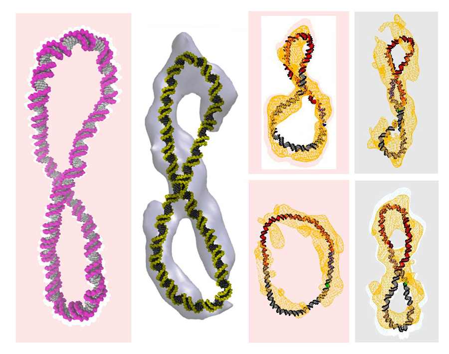
Images of the three-dimensional structure of supercoiled DNA were revealed in a study published this week in the journal Nature Communications, showing the shape is much more dynamic than the double helix developed by scientists Watson and Crick in 1978. Earlier, when Watson and Crick looked at only the tiny part of the real genome i.e. only about one turn of the double helix, which is about 12 base pairs.
There are basically three billion base pairs that holds up the entire set of DNA directions amid people.
With a goal to research the construction of DNA whereas they’re overcrowded into cells, scientists needed to replicate this coiling of DNA.
It will take a while until people will be able to digest these new DNA findings and think of DNA as a much more complicated shape that moves.
To examine how the winding altered what the circles seemed like, the researchers coiled after which uncoiled the miniscule DNA circles – which is maybe 10 million occasions shorter in size than the DNA contained inside our cells – a single flip at a time. Just like in the human body, the enzyme relaxed the twist in even the most tightly coiled DNA.
She notes, “Some of the circles had sharp bends, a few were figure-8s, and others looked like racquets or sewing needles. A few looked like rods because they were so coiled”, said Irobalieva.
Researchers from the United States and Britain are hoping to untangle this conundrum using powerful microscopes and supercomputer simulations.
“These enzymes don’t do anything to linear DNA because it’s not coiled up”, said co-author Dr. Daniel J. Catanese, Jr., also of Baylor.
While the researchers expected to see the opening of base pairs when the DNA was underwound, they were surprised to see this opening for the overwound DNA.
Typically, the DNA helix is formed when complementary base pairs – such as the nucleotide adenine and its partner guanine – bind together, forming a bridge across the helix.
Advertisement
Dr Harris concludes: “We are sure that supercomputers will play an increasingly important role in drug design. We try to do a puzzle with tens of millions of items, and so they all maintain altering form”.


